
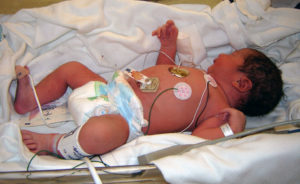
CP Caused by Periventricular Leukomalacia
There are a number of brain-related injuries that have the potential to cause cerebral palsy in newborn children. Periventricular leukomalacia (PVL) is characterized as damage to the white matter of the brain, near the lateral ventricles, and is commonly-associated with the development of cerebral palsy. In fact, it’s thought that 60 to 100 percent of children who suffer from PVL will develop cerebral palsy.
CP & Periventricular Leukomalacia
Periventricular leukomalacia involves the necrosis (death) of the brain’s white matter due to a lack of oxygenated blood reaching particular areas of the child’s brain. This type of brain injury causes empty areas to form within the brain.
The brain’s periventricular white matter is responsible for the body’s motor functions. Any damage that occurs in this area has the potential to cause spasticity, epilepsy, vision problems, and cognitive impairment.
A rather essential component of the white matter is myelin, which is responsible for insulating cell pathways and ensuring the nerve impulses transmit in a timely manner. If the myelin is damaged, nerve transmission is slowed and impeded which can also lead to impairment in brain function.
In severe cases where infants who only survived for a short period of time, studies show that about 75 percent of these newborns had suffered periventricular leukomalacia. Experts believe that infections in the uterus play a significant role in the development of PVL, as does any hypoxic (decreased oxygen flow) or ischemic (decreased blood flow) event suffered during labor or delivery.
Causes and Risk Factors
Periventricular leukomalacia can occur when there is either a reduced blood flow to the brain or damage to the cells in the periventricular tissue. The greatest risk lies with premature infants who are born before the 32nd week of pregnancy and are placed on a ventilator. There is also a risk for the development of PVL in infants who suffer from hypotension, acidosis, hypocarbia, and hypoxemia.
There are a number of factors that that increase the potential for PVL, such as the following:
- Conditions in the placental blood vessels (e.g., placental vascular anastomoses)
- Pregnancy involving twins
- Bacterial infection that causes an inflammation of the fetal membranes
- Cocaine use by the mother during gestation
- Development of sepsis
- Development of funisitis (inflammation of the connective tissue of the umbilical cord)
Diagnosis & Treatment
Babies who are at risk for the development of periventricular leukomalacia (or have already been diagnosed with PVL) will need several tests to determine whether or not serious brain damage has occurred, and to mitigate any further damage.
MRIs and cranial ultrasounds can be used to detect the presence of periventricular leukomalacia, but this isn’t always reliable if these tests are performed too early. In the event there is a suspicion of PVL, the performance of frequent developmental assessments may be recommended.
In order to manage the symptoms associated with this type of neurological injury, treatment generally focuses on the following areas: